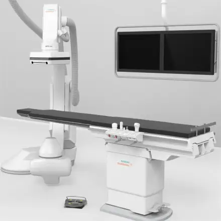
Original new Siemens syngo.CT Neuro DSA system digital subtraction angiography
Support Payment Term:

HSBC Hong Kong

paypal

Alibaba Pay

Western Union

USD

EUR

GBP

SGD

HKD

CNH

CAD

MXN

BRL

JPY

THB

MOP

AUD

NZD

PLN

CZK

HUF

RON

CHF

SEK

NOK

DKK

TRY

AED

SAR

ILS

ZAR
$0
Discover similar items
Page 1 of 2

Features & Benefits
Due to the high negative predictive value of up to 99% and low routine effective doses with the Siemens SOMATOM Definition family, the rule-out of CAD has become a safe routine procedure in many institutions. syngo.via’s dedicated automatic pre-processing immediately displays the coronary arteries in Curved Planar Reformations (CPRs) thus allowing for a rule-out of Coronary Artery Disease in less than a minute.
The Single Click Stenosis function gives you all relevant information at a glance, such as the diameter, length, and area of a stenosis, as well as the profile curve and minimum lumen identification. This allows not only for pre-surgical evaluation before intracoronary stenting or coronary bypass graft planning, but also for post-surgical evaluation for stent, bypass graft and vascular patency.
Clinical Benefits
- Automatic segmentation of the coronary tree and automatic labeling of the main coronary arteries, major coronary branches, and saphenous vein grafts
- Comprehensive layout for display of multiple CPRs permitting the review of the coronary tree with the blink of an eye. The dual monitor layout ensures an excellent overview of axial slices and volume rendered technique (VRT) images. The user may customize the screen layout according to his or her preferences.
- Calculation of the diameter, area, and length of a stenosis with the single-click stenosis function
- Enhanced lesion and stent visualization with the Image Sharpening tool. Blooming artifacts are reduced while saving the time for an additional reconstruction with a sharp reconstruction kernel.
- Robust segmentation of the coronary vessels despite high-grade stenoses
- Comprehensive 3D visualization of the coronary tree, including layered display of cardiac and coronary anatomy
Rapid Results Technology
- User-specific definition of customizable procedures that can be saved as individual protocols in the Protocol Configurator
- These configured protocols can be re-used for an automated generation of snapshots, radial and parallel ranges for MPR, MIP, and VRT images (incl. VRT presets) in every case
- Standardized image creation, including PACS series and filming, for time-saving in clinical routine
- Ideal to share hints, tips, and recommendations both for educational purposes and in order to increase standardization