
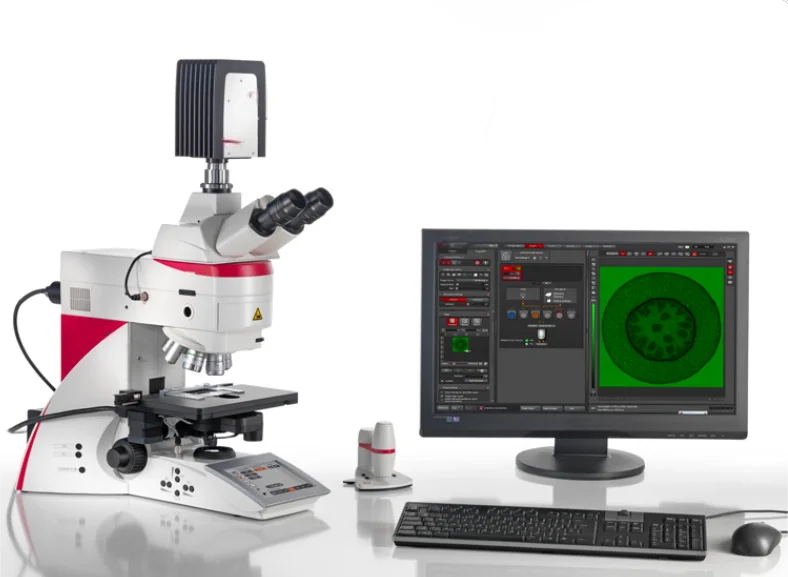
Leica DM6B Fluorescent Microscope price DM6 Bfluorescence biological microscope Fully Automated Upright Microscope System for Life Science Research
Support Payment Term:
PayPal
USD
EUR
GBP
SGD
CNY
MXN
BRL
JPY
THB
AUD
$93000.00
Discover similar items
Page 1 of 2
Specifications:
Display: Controls the microscope conveniently and intuitively
Focus: Mechanical & Motorized
Stage: Motorized (optional)
Specimen Overview Tool: Yes
Transmitted Light Illumination: LED or Halogen
Transmitted Light Contrast Methods: BF, PH, DF, POL, DIC
Fluorescence Illumination: LED or mercury metal halide
See More in Less Time
Saving time in image acquisition means you have more time for what is really at the heart of your science. Here are just a few ways to speed up your workflow.
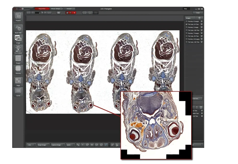
See up to 10.000x more of your sample with the LAS X Navigator. Set up high resolution image acquisition for slides, dishes and multi-well plates
Take full advantage of your sCMOS camera. The 19-mm field of view camera port perfectly fits the dimensions of common sCMOS sensors.
Make your slide examination faster at the highest resolution.
Choose the objective that best fits your application – from a range of more than 300 outstanding optics. Just one example: the unique 1.25x overview objective for a perfect wide view.
Free Your Mind!
Focus on the result you would like to achieve rather than the process of getting to it. Intelligent Automation does the work for you: It solves multi-step routines with the press of a button, makes your work life easier, saves time, and allows you to focus on your experiment, not on complex microscope settings.
Change contrast with the push of a button – everything you need automatically slides into place.
Acquire publication-ready images.
Use Light and Contrast Management to simplify your work – for example the Fluorescence Intensity Manager (FIM), fast Internal Filter wheel (IFW) and Excitation Manager (ExMan) for fluorescence images, or Koehler light management for perfect Koehler images.
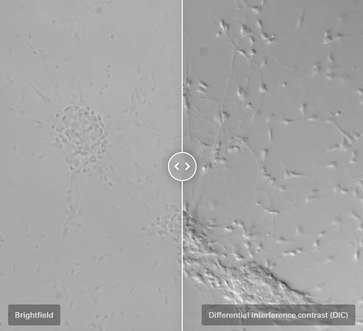
Contrast method DIC
Unstained biological specimens often do not show a very high contrast. Thick specimens in particular, such as brain slices, show up as nothing more than light grey structures instead of single cells.
Differential interference contrast
Differential interference contrast (DIC) is here an ideal contrast method.
Exactly What You Need
You can configure the microscope that fits your need and budget. Optimize your system to your application with different contrast methods and automated functions.
LED illumination lights your sample homogenously and with a constant color temperature, while also saving energy and avoiding bulb changes due to the LED’s lifetime of up to 25,000 hours
Make Your Microscope an Imaging System
Microsystems provides powerful cameras and workflow-based software to complete your integrated imaging system.
If you have any more questions, please contact us for consultation.
Trade Guarantee
Quality Assurance, Brand Supply
Specialty Selection
Rich Products,Great Variety Of Goods
Honest Service
Digital Drive, Fully Enabled
Logistics Services
Online Tracking, Fast And Reliable

 WS
WS